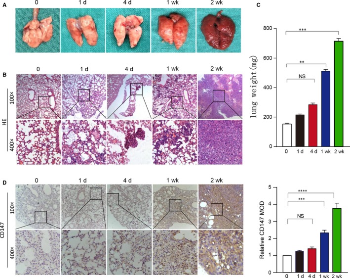
Figure 5.

CD147 expression was higher lung metastasis tissues in vivo. (A) Whole mount pictures of the lung tissues are shown. (B) H&E staining of lung tissue sections at 0, 1 day, 4 days, 1 week and 2 weeks is shown. (C) The lung weight of BALB/C nude mice killed at 0, 1 day, 4 days, 1 week and 2 weeks are shown. ****P < 0.0001, based on Student's t test. (D) Immunohistochemical staining of CD147 in lung tissue sections at 0, 1 day, 4 days, 1 week and 2 weeks is shown. ****P < 0.0001, based on Student's t test
