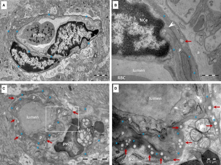
Figure 2.

TEM of brain capillaries in a 6‐month‐old mouse (A, B) and a 24‐month‐old mouse (C, D). A, A capillary from the 6‐month‐old mouse has a uniform BM (*) subjacent to an endothelial cell (NCe—nucleus of the endothelial cell; e—endothelial cell) and enclosing a pericyte (p). PMNn—polymorphonuclear neutrophil; a—end‐feet of astrocytes. B, Rare droplets (arrow) may be seen in the BM (*) of capillaries from the young mouse brain. Arrowhead indicates a direct contact between a pericyte and an endothelial cell. C, The BM (*) of brain capillaries from the aged mouse is thicker, uneven and contains numerous electron‐lucent, single or grouped droplets (arrows). Large lipid‐containing lysosomes (ly) are present in a perivascular cell (pvc). p –pericyte; a—end‐foot of astrocyte. D, Higher magnification of marked area in C
