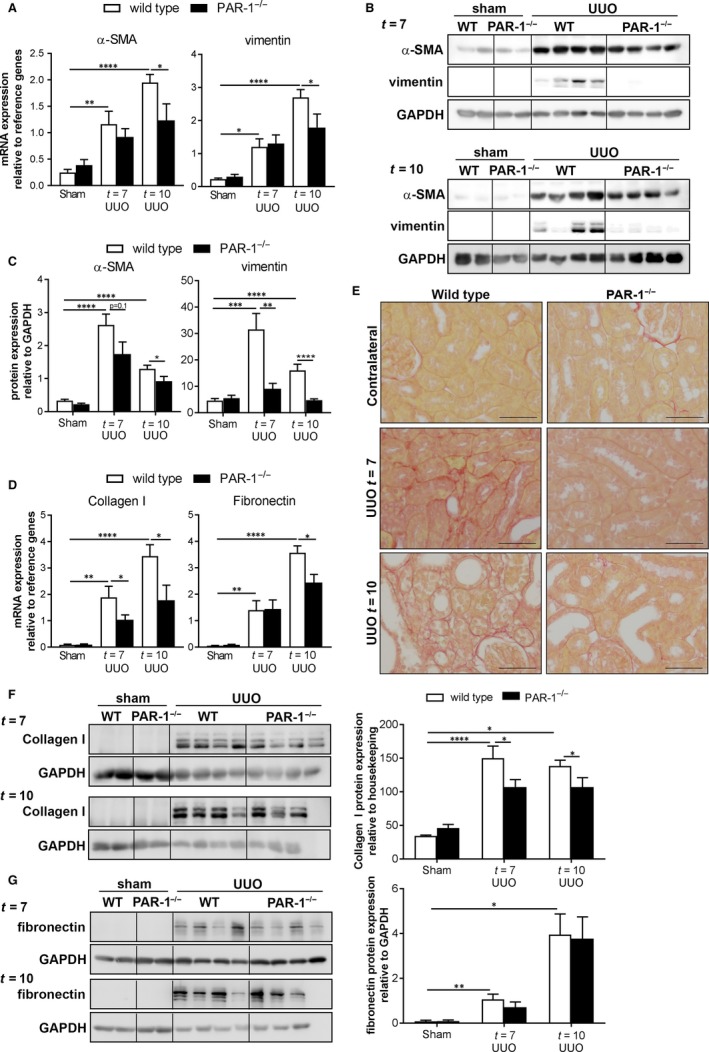
Figure 3.

PAR‐1 deficiency limits renal fibrosis. A, α‐SMA and vimentin mRNA expression in whole kidney lysates of unobstructed (sham) and obstructed (UUO) kidneys of wild‐type (WT) and PAR‐1‐deficient (PAR‐1−/−) mice, 7 and 10 d after UUO. B, Western blot analysis of α‐SMA and vimentin in whole kidney lysates of unobstructed (sham) and obstructed (UUO) kidneys of wild‐type (WT) and PAR‐1‐deficient (PAR‐1−/−) mice, 7 and 10 d after UUO. GAPDH expression served as loading control. C, Quantification of Western blots depicted in panel B. D, mRNA expression of collagen I and fibronectin in whole kidney lysates of unobstructed (sham) and obstructed (UUO) kidneys of wild‐type (WT) and PAR‐1‐deficient (PAR‐1−/−) mice, 7 and 10 d after UUO. E, Representative pictures of picrosirius red staining. F‐G, Western blot analysis (left: representative picture, right: quantification) of collagen I (F) and fibronectin (G) in whole kidney lysates of unobstructed (sham) and obstructed (UUO) kidneys of wild‐type (WT) and PAR‐1‐deficient (PAR‐1−/−) mice, 7 and 10 d after UUO. GAPDH expression served as loading control. *P < 0.05; **P < 0.01; ***P < 0.005; ****P < 0.001 (one‐way ANOVA followed by Bonferroni multiple comparisons test)
