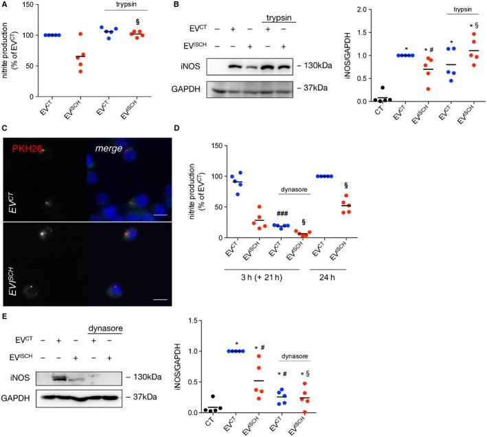
Figure 2.

Vesicle surface proteins are implicated in macrophage activation induced by EVs from ischaemic cardiomyocytes. (A) EVCT or EVISCH were treated with 1 mg/mL trypsin for 10 minutes. Trypsin‐treated or untreated EVs were further incubated with macrophages for 24 hours. Nitrite production was determined and results are expressed as percentage of nitrite production over macrophages treated with EVCT. § P < 0.05 vs EVISCH (n = 4). (B) iNOS expression was evaluated in macrophages challenged with trypsin‐treated or untreated EVCT or EVISCH for 24 hours, by WB. Graph depicts quantification levels. *P < 0.05 vs CT, # P < 0.05 vs EVCT, § P < 0.05 vs EVISCH (n = 5). (C) WGA‐stained macrophages (green) were incubated with PKH26‐labelled EVCT or EVISCH (red) for 30 minutes, after which cells were fixed and visualized by fluorescence microscopy. Nuclei were stained with DAPI (blue). Scale bars 5 μm. (D) Macrophages were treated with 80 μmol/L dynasore for 30 minutes, followed by incubation with EVCT or EVISCH for 3 hours. Medium was then replaced with EV‐depleted medium and cells were kept in culture for an additional 21 hours. Nitrite production was determined and results are expressed as percentage of nitrite production over macrophages treated with EVCT for 24 hours. Cells challenged with EVCT or EVISCH for 24 hours were used as control. ### P < 0.001 vs EVCT, § P < 0.05 vs EVISCH (n = 5). (E) iNOS expression was evaluated in macrophages either treated or not with dynasore, followed by stimulation with EVCT or EVISCH, by WB. Graph depicts quantification levels. *P < 0.05 vs CT, # P < 0.05 vs EVCT, § P < 0.05 vs EVISCH (n = 5)
