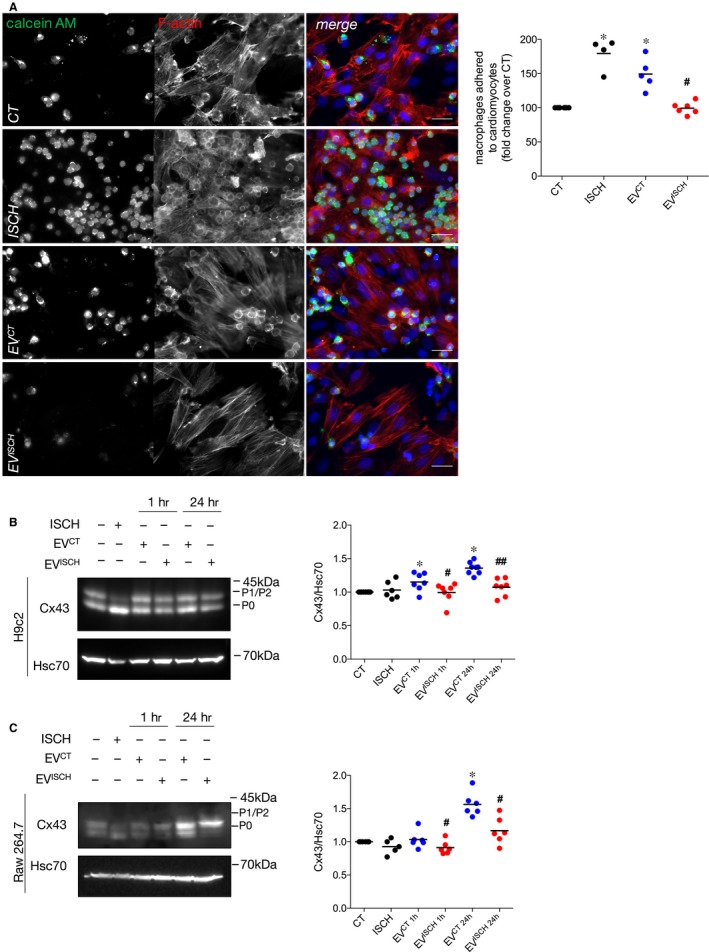
Figure 3.

Control EVs increase macrophage adhesion to cardiomyocytes. (A) Macrophages were labelled with 1.5 μmol/L calcein‐AM (green) and further incubated with previously adherent H9c2 cells for 1 hour. Where indicated, EVCT or EVISCH were added or simulated ischaemia was performed for 1 hour. F‐actin was stained with Rhodamine‐Phalloidin (red), and nuclei were stained with DAPI (blue). Scale bars 20 μm. Graph depicts the number of adherent macrophages. *P < 0.05 vs CT, # P < 0.05 vs EVCT (n = 4‐6). (B) H9c2 cells were incubated with EVCT or EVISCH for 1 or 24 hours. Cx43 levels were analysed by WB. Hsc70 was used as loading control. Graph depicts normalized levels of Cx43. *P < 0.05 vs CT, # P < 0.05, ## P < 0.01 vs EVCT (n = 6‐7). (C) Raw 264.7 cells were incubated with EVCT or EVISCH for 1 or 24 hours. Graph depicts normalized levels of Cx43. *P < 0.05 vs CT, # P < 0.05 vs EVCT (n = 5‐6)
