Figure 4.

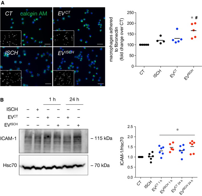
Ischaemic EVs increase adhesion of macrophages to a fibronectin matrix. (A) Macrophages were labelled with 1.5 μmol/L Calcein‐AM (green) and added on top of a fibronectin‐based matrix. Where indicated, EVCT or EVISCH were added or simulated ischaemia was performed for 1 hour. Nuclei were stained with DAPI (blue). Scale bars 20 μm. Graph depicts the number of adherent macrophages. *P < 0.05 vs CT, # P < 0.05 vs EVCT (n = 4‐5). (B) Macrophages were incubated with cardiomyocyte‐derived EVCT or EVISCH for 1 or 24 hours. Graph depicts the levels of ICAM‐1. *P < 0.05 vs CT (n = 5‐6)
