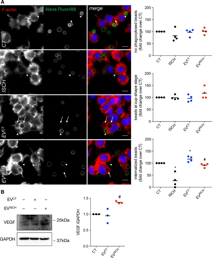
Figure 5.

Control EVs increase the phagocytic capacity of macrophages. (A) Macrophages were incubated with opsonized latex beads for 20 minutes (ratio of 10 beads/cell). Non‐internalized beads were labelled with Alexa Fluor 488 antibodies (green). F‐actin was stained with Rhodamine‐Phalloidin (red). Graphs depict the total number of beads/phagocytic macrophages, represented as percentage of fold change over control (CT), the number of beads within cup‐shaped unsealed nascent phagosomes (positive Alexa Fluor 488 staining, arrow heads) and the number of beads in sealed phagosomes (negative Alexa Fluor 488 staining, arrows). Nuclei were stained with DAPI (blue). Scale bars 10 μm. *P < 0.05 vs CT, # P < 0.05 vs EVCT (n = 4).(B) Macrophages were incubated with EVCT or EVISCH, for 24 hours. Graph depicts quantification of VEGF levels analysed by WB. # P < 0.05 vs EVCT (n = 3)
