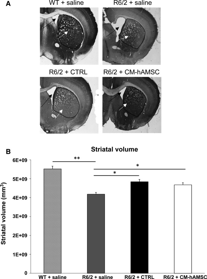
Figure 3.

Effects of Conditioned medium from hAMSC (CM‐hAMSC) treatment on striatal atrophy in R6/2 mice at 13 weeks of age. A, Transmitted light microscope images showing representative calbindin‐stained coronal sections of a wild‐type (WT) mice treated with saline, and saline‐, CTRL‐ or CM‐hAMSC‐treated R6/2 mice. Marked gross striatal atrophy (black arrows) and enlarged lateral ventricles (white arrows) are present in vehicle‐treated R6/2 mice compared to wild‐type mice. This atrophy is mostly absent from the sections of the R6/2 mouse treated with CTRL or CM‐hAMSC. B, Quantification of differences in striatal volume in the same groups as (A). Post hoc analysis indicated that R6/2 mice treated with saline had a significantly reduced striatal volume compared to the wild‐type group (*P < 0.05; **P < 0.01)
