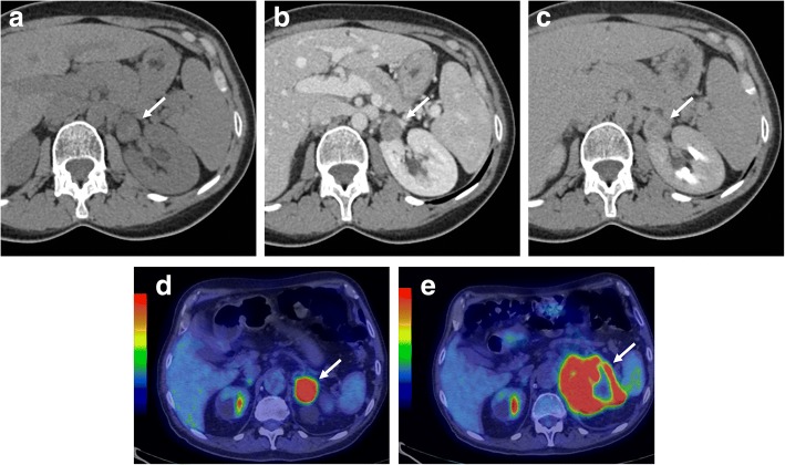
Fig. 10.
Left adrenal metastasis in a 62-year-old woman with lung cancer. Unenhanced CT (a), portal (b), and late phase (c) images show a left adrenal mass (arrow) with attenuation value of 26 HU on unenhanced CT (a) and “absolute percentage washout” of 15%. Staging 18F-FDG PET/CT (d) performed 3 months later revealed a left adrenal lesion (arrow) with high metabolic activity (SUVmax 14 g/mL) and increased in size in comparison with CT images. After chemotherapy, restaging 18F-FDG-PET/CT (e) showed morpho-functional disease progression of the adrenal lesion (arrow) with signs of central necrosis (SUVmax 19 g/mL with central necrotic area of absence of 18F-FDG uptake). Images of 18F-FDG-PET/CT provided by database of Nuclear Medicine Service, Fondazione Istituto G. Giglio, Cefalù, Italy

