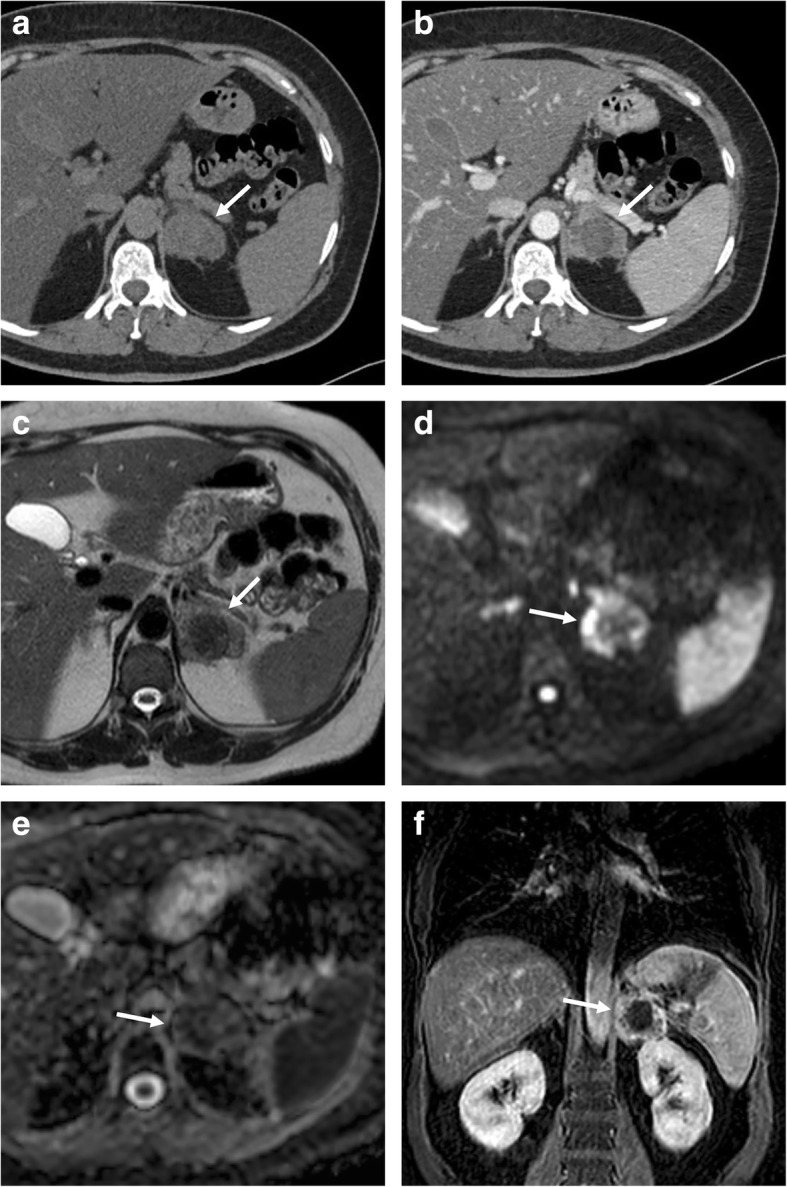
Fig. 11.

Left adrenal metastasis in a 58-year-old woman with breast cancer. Axial unenhanced CT (a), portal phase (b), T2-weighted fast spin echo (c), b800 s/mm2 diffusion-weighted image (d), ADC map (e), and coronal three-dimensional fat-suppressed T1-weighted gradient recalled echo image in portal phase after contrast injection (f) show a left adrenal mass (arrow). This lesion intra-lesional hemorrhage presenting as spontaneously hyperdense area on unenhanced CT (a), hypointense on T2-weighted (c), and no enhancement on CT (b) and MR (f) images after contrast injection. The peripheral solid component of the lesion shows also restricted pattern of diffusion (d, e)
