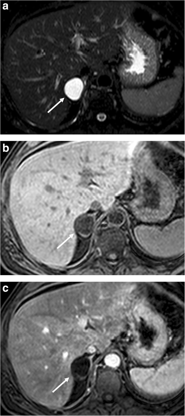
Fig. 4.

Right adrenal cyst in a 50-year-old woman. Axial T2-weighted fat-saturated fast spin echo MR image (a), three-dimensional fat-suppressed T1-weighted gradient recalled echo images before (b) and after paramagnetic contrast administration (c) show a right adrenal cystic lesion (arrow) with low T1 signal intensity, high T2 signal intensity, no contrast enhancement and no septations, blood products, soft-tissue components, or calcifications
