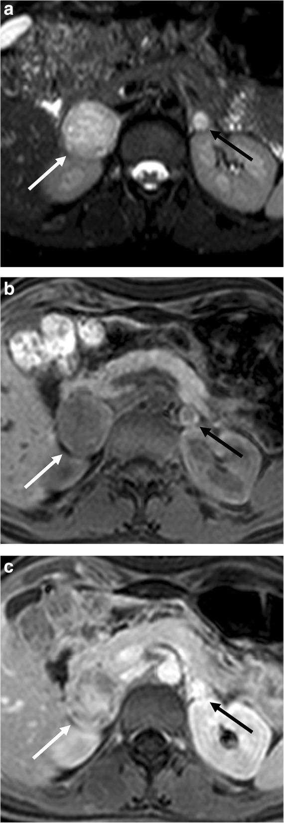
Fig. 6.

Bilateral adrenal pheochromocytoma in a 27-year-old woman with multiple endocrine neoplasia type 2. Axial T2-weighted fat-saturated spin echo (a), three-dimensional fat-suppressed T1-weighted gradient recalled echo images before contrast injection (b), and in portal phase (c) images show a bilateral adrenal mass with high T2 signal intensity and strong and heterogenous contrast enhancement. The signal intensity is typically more inhomogenous in larger lesions (white arrow, right adrenal gland) than in smaller ones (black arrow, left adrenal gland)
