Figure 1.

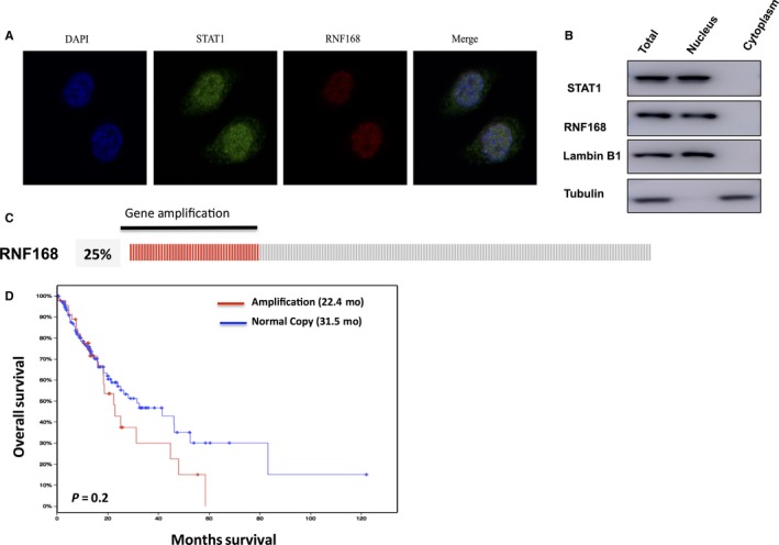
RNF168 has high frequency of gene amplification in oesophageal cancer and tends to correlate with poor prognosis in oesophageal cancer patients. (A) NEC cells cultured on sterile glass cover slips were fixed with 4% formaldehyde for 10 min. Then the cells were incubated in permeabilization buffer (0.3% Triton X100, in phosphate buffered saline [PBS]) for 10 min. Ten percent donkey serum was added to suppress nonspecific antibody binding at room temperature for 30 min and primary antibodies were incubated overnight at 4°. Fluorochrome‐conjugated secondary antibodies were added after wash in a dark chamber at room temperature. The slides were washed with PBS and mounted using mounting solution containing DAPI. Finally, slides were visualized with a NIKON80i fluorescent microscope. The antibodies were used as follows: anti‐RNF168 (SC‐101125, santa cruz); anti‐STAT1 (9172, Cell Signalling Technology). (B) RNF168 is mainly localized in the nuclear. The subcellular protein fractionation kit (Thermo Scientific, 78840) was used for cytoplasm and nuclear separation. Tubulin and Lambin B1 were used for cytoplasm and nuclear control. (C, D) RNF168 has gene amplification in 25% of esophageal cancer patients. The TCGA database was used to extract the gene expression data. RNF168 gene amplification tends to correlate with poor overall survival in esophageal cancer patients
