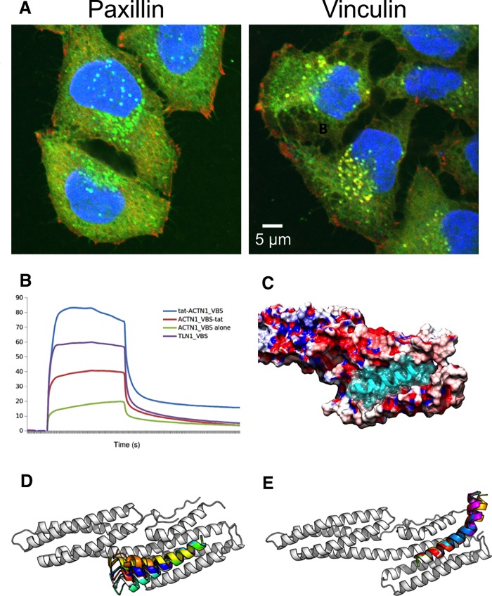
Fig 3. Specificity of vinculin binding region peptides.
(A) HeLa cells treated with 5FAM-labeled tat-ACTN1-VBS peptide and immunostained with either vinculin (right) or paxillin (left) antibody. Blue: nuclear stain; green: tat-ACTN1-VBS peptide, conjugated with the 5FAM fluorophore at the N terminus; red: paxillin or vinculin staining, as labeled; yellow: co-localization of protein and peptide. Full results in S10 Fig. (B) Surface plasmon resonance sensorgrams for serial peptide injections at 1 mM onto immobilised vinculin on a flow cell of a CM5 sensor chip. Relative binding affinity tat-ACTN1_VBS > TLN1_VBS-tat > ACTN1_VBS-tat > ACTN1_VBS. (C) Electrostatics of the vinculin surface: Electrostatic surface showing the active site of the alpha actinin peptide ACTN1-VBS (not including tat) binding to vinculin (PDB entry 1YDI). The region in which the positively charged tat sequence is likely to bind is a negatively charged (red) region of vinculin. Positively charged regions are shown in blue. (D) FlexPepDock [28] was used to predict the binding poses of the ACTN1-VBS-tat peptide where the tat peptide is located at the peptide C-terminus, to the PDB entry 1YDI, contrasting alternative termini for coupling with tat. (E) As for (D), but with the more stably binding tat-ACTN1-VBS peptide, in which the tat region at the N-terminus of the peptide (top right of image) appears to have been stabilized in comparison with (D), via interactions of the positively charged tat peptide with a negatively charged surface region of vinculin.

