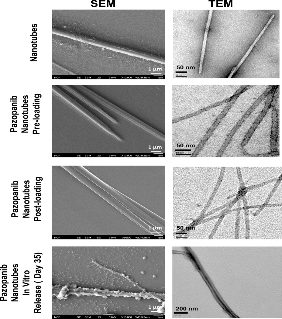
Figure 4.
Scanning electron microscopy (SEM) (left panel) and transmission electron microscopy (TEM) images (right panel) showing surface morphology and particle size of plain, pazopanib loaded nanotubes and pazopanib loaded nanotube samples after 35 days of in vitro release. Pictures were taken using a scanning electron microscope at magnifications ×10,000 and using transmission electron microscope. TEM images of drug loaded tubes had a dense interior compared to free dipeptide nanotubes that have transparent core, suggesting positive drug loading. Samples isolated from 35 days in vitro release media showed shrunken and ridged surface as compared to plain and freshly drug loaded nanotubes, suggesting nanotube surface erosion in the acetate buffer release media (pH 4.5) as one possible drug release mechanism along with simple diffusion.

