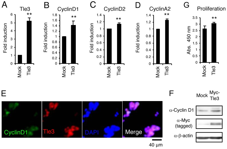
Figure 2. Overexpression of Tle3 increases proliferation in B16 melanoma cells.
(A-F) B16 cells stably expressing Myc-tagged Tle3 or empty vector were generated after positive selection with G418. The messenger RNA levels of Tle3 (A), CyclinD1 (B), CyclinD2 (C), or CyclinA2 (D) were determined by qPCR on day 2. B16 cells with high expression of Tle3 co-expressed CyclinD1 in the nuclei. Scale bar corresponds to 40 μm (E). Protein levels of CyclinD1, Myc-tagged Tle3, or β-actin were determined by western-blot analysis on day 2 (F). Overexpression of Tle3 increased the proliferation of B16 cells assessed by water-soluble tetrazolium salt (WST) -8 assay. Proliferation was quantified on day 2 by spectrophotometric absorbance measurement at 450 nm (G). Data are expressed as the mean ± SD (n = 3). **, p < 0.01 versus control (A-D, G). Representative images were shown (E and F).

