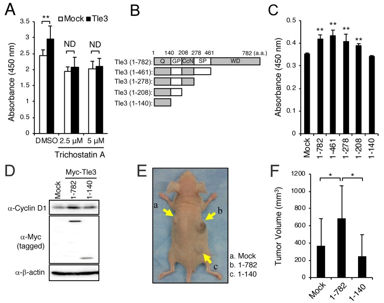
Figure 6. HDACs are involved in the enhancement of the proliferation of B16 cells by Tle3.
(A-C) B16 cells were transiently transfected with empty vector (Mock) or Myc-tagged Tle3 and then treated with DMSO, or the indicated concentration of trichostatin A. Cell proliferation was evaluated on day 2 by water-soluble tetrazolium salt (WST) assay and absorbance measurement at 450 nm (A). Schematic of the C-terminally truncated forms of the Myc-tagged Tle3 plasmids used in these experiments. Q; glutamine rich domain, GP; glycine/proline rich domain, CcN; CcN domain, SP; serine/proline rich domain, WD, tryptophan/aspartic acid repeat domain (B). C-terminally truncated forms of Tle3 were transfected in B16 cells and proliferation ability measured on day 2 by WST assay (C). The data are expressed as the mean ± SD (n = 3). **, p < 0.01 versus Mock transfection (A and C). B16 cells were transfected with empty vector (Mock), Myc-tagged Tle3 (1-782), or Myc-tagged Tle3 (1-140). Protein levels of cyclinD1, Myc (tagged) or β-actin were assessed by western blotting analysis on day 2 (D). (E and F) B16 cells stably expressing Myc-tagged Tle3 (1-782), Myc-tagged Tle3 (1-140), or empty vector were generated after positive selection with G418. BALB/cA Jcl-nu/nu mice (n=5) were injected subcutaneously with 1 × 10⁵ mock B16 cells (a), cells stably expressing Myc-tagged Tle3 (1-782) (b), or cells stably expressing Myc-tagged Tle3 (1-140) (c). Representative photograph of a mouse 3 weeks after injection with B16 cells (E). The volume of resected tumors was quantified. The data are expressed as the mean ± SD (n = 5). *, p < 0.05 (F).

