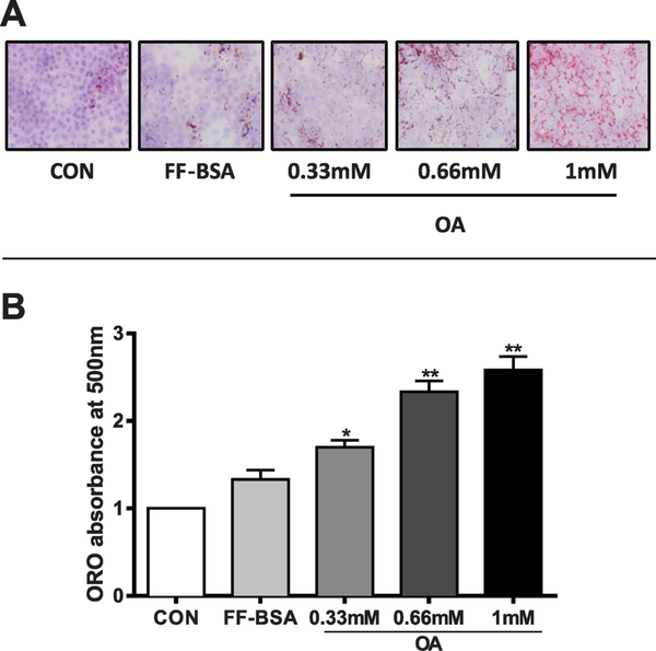
Figure 1. Determination of neutral lipid accumulation by ORO staining in OA-loaded HepaRG cells.
HepaRG cells were incubated for 48 hours in HepaRG treatment medium alone (CON) or containing 0.7% FF-BSA, either alone or complexed with 0.33 mM, 0.66 mM, or 1 mM OA. Treatment medium was replaced once after 24 hours. (A) ORO was used to stain neutral lipids, and cells were photographed under phase-contrast optics at 200X magnification. Neutral lipids are stained red, hepatocyte nuclei are purple/blue. (B) ORO was extracted from the cells and absorbance measured at 510 nm. Each bar represents the mean ± SEM (n=4 wells per group, derived from combining the data from 2 independent HepaRG experiments with duplicate treatments). *Significantly different from vehicle control (FF-BSA), P<0.05, **P<0.01.

