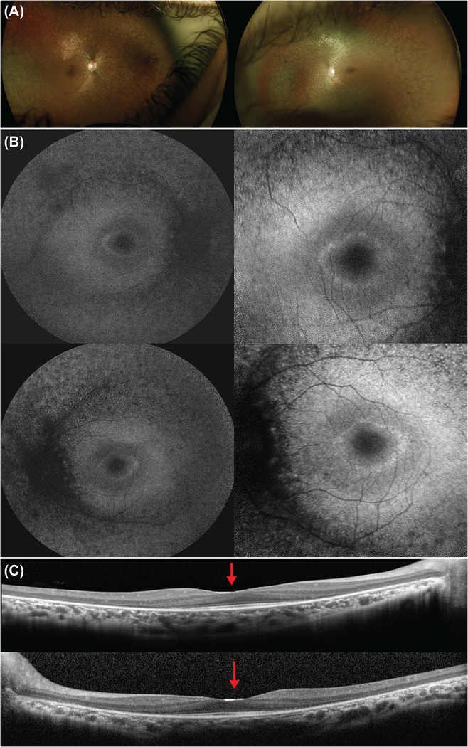
Figure 2. Multimodal imaging of the proband at presentation.
(a) Color fundus images of the right and left eye, respectively, showed attenuated vessels, pale optic disc, peripheral retina atrophy, and bone-spicule intraretinal pigment migration. (b) Fundus autofluorescence images of the right (top) and left (bottom) eye demonstrated peripheral atrophy and the presence of a hyperautofluorescent ring on the foveal area. The ring is seen in more detail with the 30-degree images (right column). (c) Spectral-domain optical coherence tomography scan through the fovea revealed peripheral thinning of the retina. The ellipsoid zone was disrupted peripherally and conserved in the foveal area. In addition, the foveal border is enlarged and flattened, with a shallow foveal pit (red arrow).

