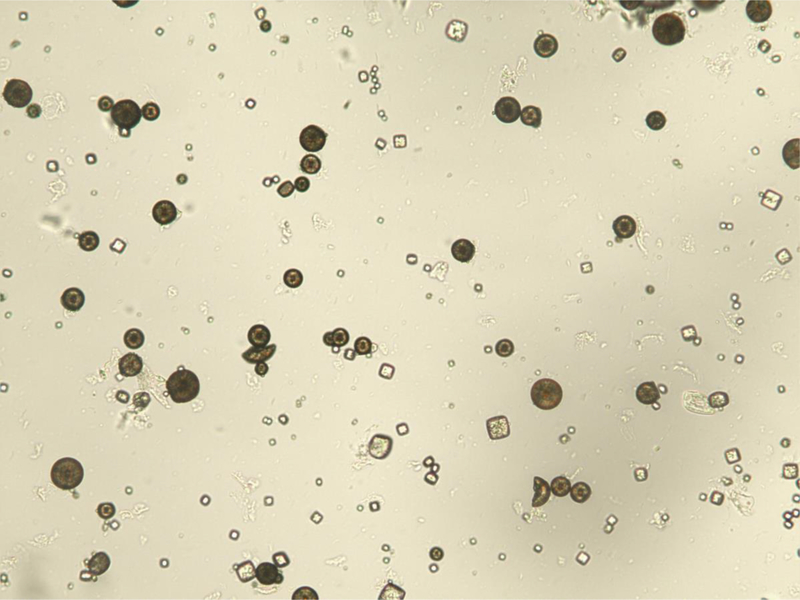
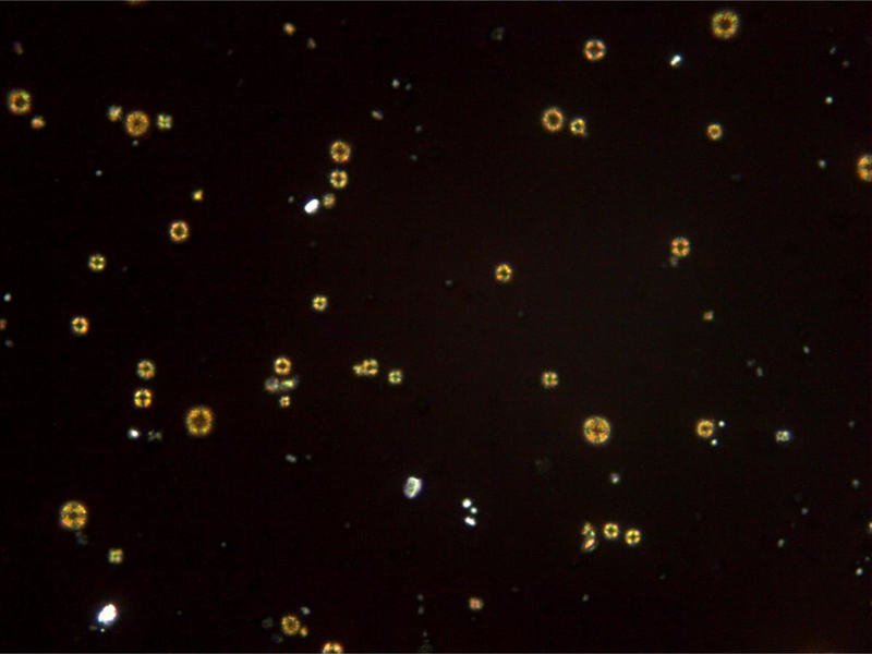
Figure 1.
Urinary 2,8-dihydroxyadenine crystals. (A) The characteristic medium-sized crystals are brown with a dark outline and central spicules. (Original magnification x 400). (B) The same field viewed with polarized light microscopy shows that the small- and medium-sized crystals appear yellow in color and produce a central Maltese cross pattern. (Original magnification x 400)


