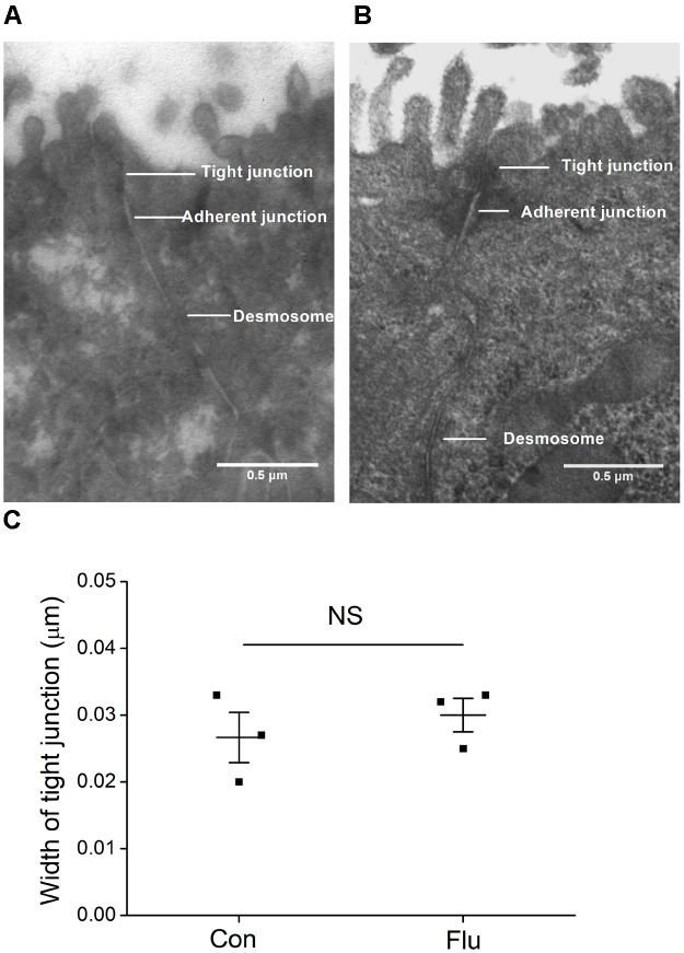
FIGURE 2.
Ultrastructure in MTECs by transmission electron microscope. (A) Tight junctions in uninfected MTECs (Con), which include tight junctions, adherent junctions and desmosome. (B) Tight junctions in influenza virus infected (Flu) MTECs. (C) Tight junction width in MTECs, NS, P > 0.05, n = 3. Data was presented as mean ± SE Student’s t-test was used to analyze the difference of the means for significance.

