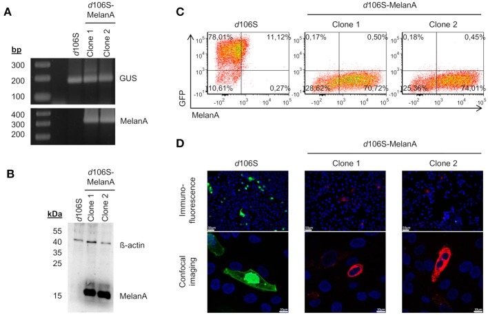
Figure 2.
Characterization of HSV-1 d106S-MelanA. MelanA expression in E11 cells infected with HSV-1 d106S and two non-fluorescent viral clones (MOI 10), which were obtained after cotransfection using HSV-1 d106S DNA and transfer plasmid pd27B-MelanA, as evident from (A) PCR, (B) Western blot, and (C) flow cytometry. Controls were (A) housekeeping gene ß-glucuronidase (GUS) and (B) ß-actin protein. (D) Expression of GFP and MelanA in Vero cells infected with HSV-1 d106S and two non-fluorescent HSV-1 d106S-MelanA clones, analyzed using immunofluorescence microscopy (upper panel) and confocal imaging (lower panel). The immunofluorescence and confocal images were taken using the DMI 6000B inverted microscope (20 × magnification) and the TCS SP5 laser scanning microscope (40 × magnification, 2.5 × zoom), respectively. Scale bars represent 50 and 10 μm in immunofluorescence and confocal images, respectively.

