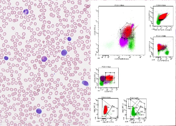
Fig. 1.
Left: Blood smear showing plasma cells constituting > 20% of total leukocytes. The plasma cells are atypical with high nuclear/cytoplasmic ratio. Right: flow cytometry histograms of blood. The neoplastic plasma cells indicated in red and purple (CD56 positive and negative fraction respective) express CD138, bright CD38, CD45, cytoplasmic kappa and are negative for CD19 and cytoplasmic lambda

