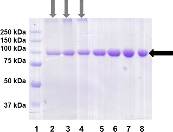
Figure 3.

Purified recombinant fusion proteins (after transient Expi293 transfection, protein A affinity chromatography and desalting) were analysed by SDS/PAGE under non-reducing conditions. Five µg of protein were applied to each lane. Several independent batches of each protein were tested (IL-2/Fc in lanes 2–4 and K35E IL-2/Fc in lanes 5–8). Black arrow indicates the major band corresponding to the fusion protein homodimer. Grey arrows highlight the presence of denaturation-resistant aggregates with lower electrophoretic mobility in all samples of non-mutated IL-2/Fc.
