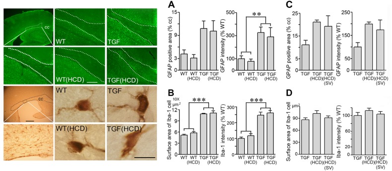
Fig. 4. Effects of high cholesterol diet (HCD) and simvastatin (SV) on white matter astrgliosis and microgliosis.
The surface area occupied by, and intensity of, GFAP-positive astrocytes (green, Cy2) were increased in the corpus callosum (cc) of TGF mice and HCD-treated TGF mice compared with WT controls, as shown here in aged TGF mice (a) (n = 5–6 mice/group). Similarly, Iba-1-immunopositive microglial cells (DAB, brown) in aged TGF mice displayed an active phenotype with increased cell body and proximal processes surface area (b). In adult TGF mice fed a HCD, SV had no effect on either phenotype (c, d) (adult, n = 4 mice/group). The area of the cc used for quantitative analysis is shown by the dotted lines. Bars: 20 μm (GFAP staining); 500 μm (Iba-1 staining in top left panel); 10 μm (single Iba-1 positive microglial cells). **p < 0.01; ***p < 0.001

