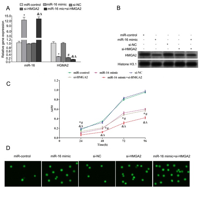
Figure 3.
Overexpression of miR-16 promotes apoptosis but inhibits proliferation of HP75 cells. (A) The expression levels of miR-16 and HMGA2 in HP75 cells following different transfections [miR-control (scramble), miR-16 mimic, si-NC, si-HMGA2 and miR-16 mimic+si-HMGA2 transfection groups] were assessed using reverse transcription-quantitative polymerase chain reaction. (B) The expression of HMGA2 was assessed using western blot analysis. Histone H3 was used as a nuclear loading control. (C) Cell proliferation was assessed using Cell Counting Kit-8. (D) Apoptosis was assessed using TdT UTP nick end labeling assay. The cells with green fluorescence were defined as apoptotic cells. *P<0.05 vs. miR-control; #P<0.05 vs. si-NC; &P<0.05 vs. miR-16 mimic+ si-HMGA2 vs. miR-control; ΔP<0.05 vs. si-NC. miR, microRNA; HMGA2, high mobility group A2; si, small interfering RNA; NC, negative control.

