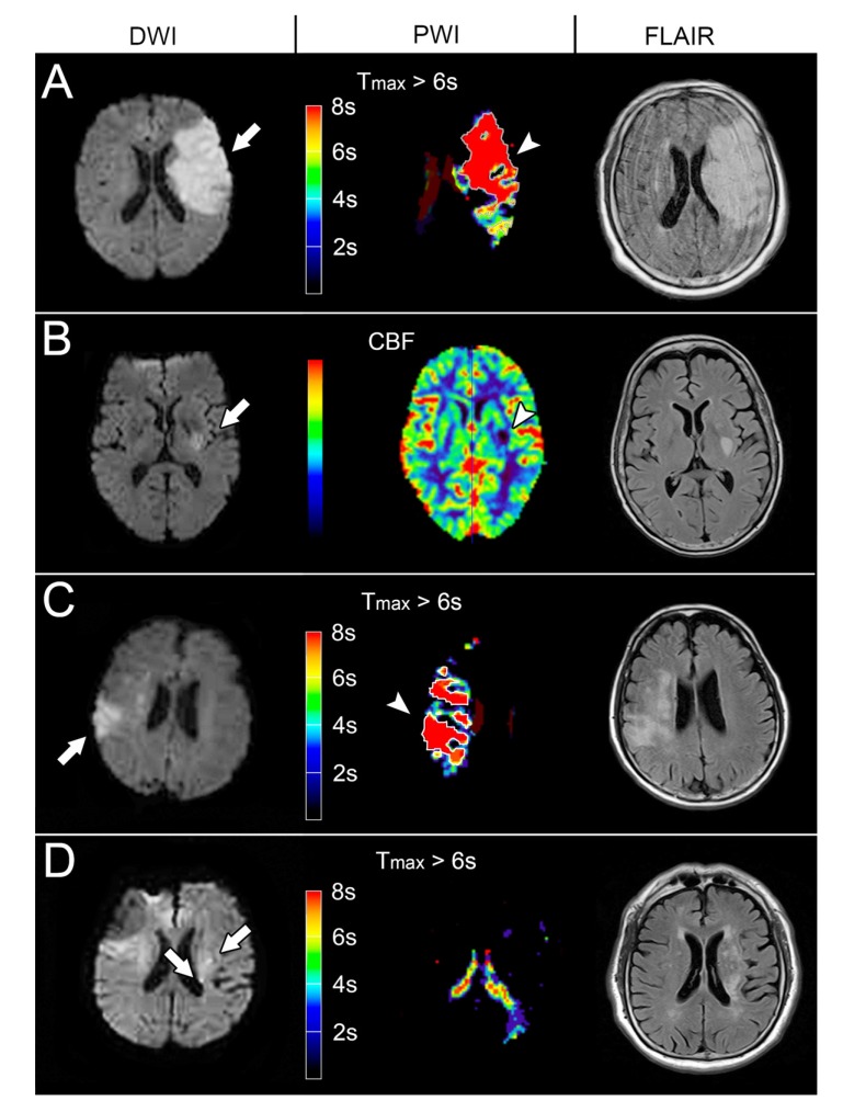
Fig. (3).
The END models of the representative patients with Diffusion Weighted Imaging (DWI), Perfusion-Weighted Image (PWI) and Fluid-Attenuated Inversion Recovery (FLAIR) imaging. (A) The DWI at 19.7 hours after stroke onset showed an acute infarct in the left Middle Cerebral Artery (MCA) territory (97.2ml, arrow), indicating positive infarct swelling model. The follow-up FLAIR imaging showed progressed infarct and brain swelling (119.8ml). (B) The DWI at 7.6 hours after stroke onset showed an acute infarct in the posterior limb of the left internal capsule (3.5ml, arrow) and a visible perfusion defect in the CBF maps (arrowhead), representing positive small subcortical infarct model. The follow-up FLAIR imaging showed infarct growth (9.3ml). (C) The DWI at 8.2 hours after stroke onset showed an infarction in the right MCA territory (22.6ml, arrow) and larger perfusion defect (Tmax > 6 seconds: 37.3 ml; arrowhead). The Tmax > 6s /infarct core ratio was 1.65 suggesting a mismatch model. The follow-up FLAIR imaging showed infarct growth (31.5 ml). (D) The DWI at 9.0 hours showed multiple acute infarction (0.6 ml, arrow) in the left centrum semiovale without perfusion defect, suggesting a recurrence model. The follow-up FLAIR imaging showed infarct growth (9.0 ml).

