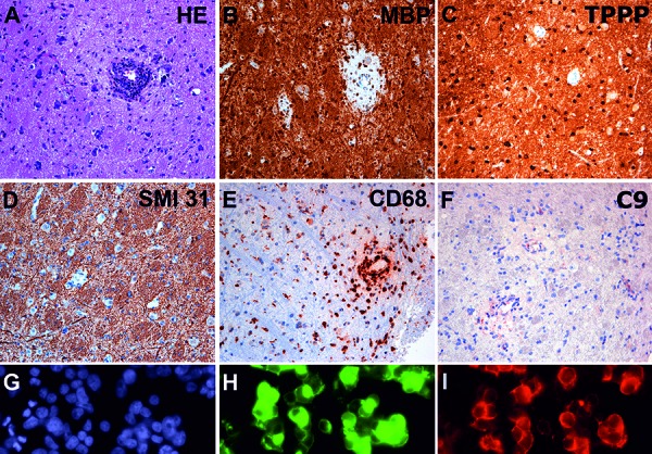
Figure 1. A: Hematoxylin & eosin (H & E) stained section shows CNS tissue with increased cellularity due to reactive astrocytes. A dense perivascular inflammatory infiltrate is observed in the center of the image. There is a slight perivascular pallor of the tissue. B: Immunohistochemistry for myelin basic protein (MBP) reveals prominent perivascular demyelination, while C: TPPP positive oligodendrocytes are well preserved. D: Immunohistochemistry for phosphorylated neurofilament SMI 31 of the same spot shows preservation of axons, confirming the selective demyelinating nature of the lesion. E: Immunohistochemistry for CD68 shows abundant activated microglia and macrophages that concentrate around the vessel. F: Mild perivascular complement deposits in the lesions (C9). G, H and I represent a cell-based assay for anti-MOG antibodies on HEK 293T cells. G (blue) represents nuclear staining, H (green) represents the effective transfection of the cells with the full-length MOG C-terminally fused with EGFP and I (red) shows the labeling of MOG-transfected HEK cells with the patient´s serum, confirming the presence of anti-MOG antibodies.

