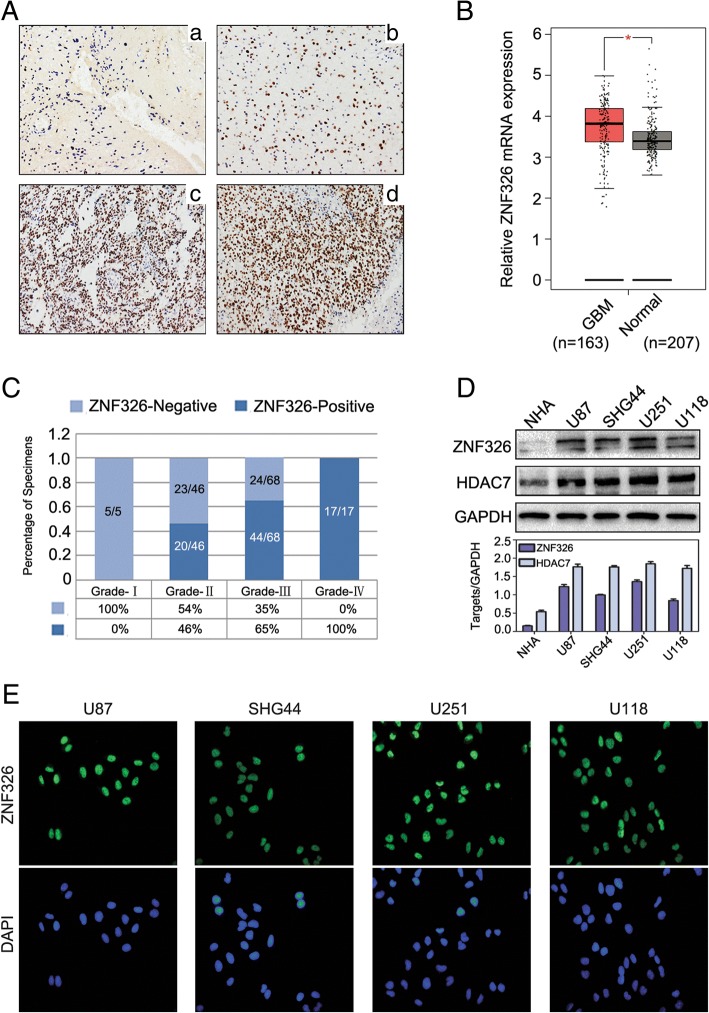
Fig. 1.
Expression and localisation of ZNF326 in glioma tissues and cell lines. a ZNF326 was negative in pilocytic astrocytoma, ZNF326-positive nuclear staining percent/HPF: < 1%, GradeI, (A-a, 400×), and positive nuclear expression (+) in diffuse astrocytoma, ZNF326-positive nuclear staining percent/HPF: 25%, grade II, (A-b, 400×); strongly positive expression (++ to +++) in anaplasia astrocytoma, ZNF326-positive nuclear staining percent/HPF: 75%, grade III (A-c, 400×) and glioblastoma, ZNF326-positive nuclear staining percent/HPF: > 90%, grade IV,(A-d, 400×). b ZNF326 mRNA expression in glioma and normal brain tissues analysed by TCGA database (P < 0.05). c The statistical view of positive expression of ZNF326 in gliomas and the positive staining percentage in different grades. d ZNF326 and HDAC7 expression was detected in a panel of four glioma cell lines and normal human astrocyte (NHA), using immunoblotting, GAPDH served as a loading control. e Immunofluorescence showed the expression and subcellular localization of ZNF326

