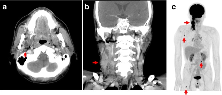
Fig. 1.
Patient computed tomography scan and positron emission tomography/computed tomography images. a and (b) Computed tomography showed a mass in the right buccal mucosa (red arrow) that extended superiorly to destruct the lateral wall of the maxillary sinus, inferiorly to the retromolar trigone, and laterally to the buccinator muscle and the anterior border of the masseter muscles, with multiple cervical lymph node enlargement. c Whole-body 18F-fludeoxyglucose positron emission tomography/computed tomography showed increased uptake in multiple lymph nodes in the right cervical area, right scapula and erector spinae muscles, and right femur (red arrows)

