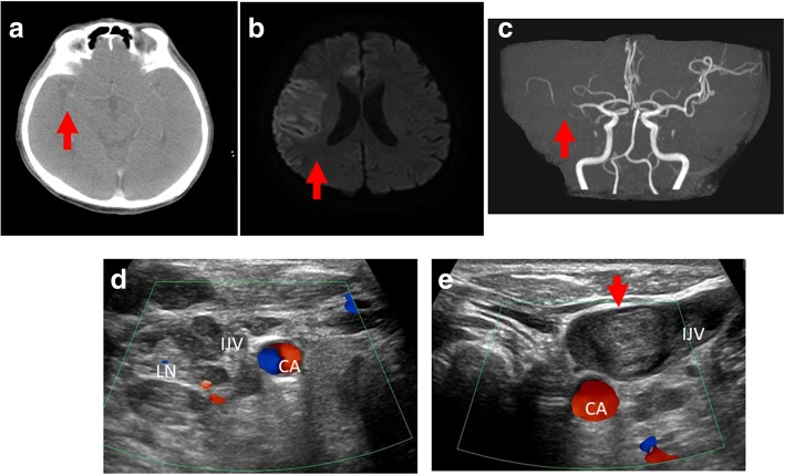
Fig. 2.
Patient computed tomography scan images after onset of aphasia and loss of consciousness. a Scattered hyperdense curvilinear areas (red arrow) suggestive of developing petechial hemorrhage in the region of the right middle cerebral artery. b Diffusion-weighted image showed a scattered lesion (red arrow) affecting the cortical part supplied by the right middle cerebra artery with corresponding deficit. c Head magnetic resonance angiography showed attenuated flow-related signal in middle cerebral artery beyond the M1 segment, while its superior division was not visible (red arrow). d A Doppler ultrasound scan of the neck revealed that the right internal jugular vein was compressed by metastatic lymph nodes. e A thrombosis was detected in the left internal jugular vein (red arrow). CA carotid artery, IJV internal jugular vein, LN metastatic lymph node

