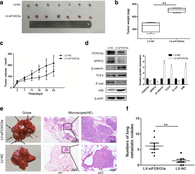
Fig. 12.
FOXO3a knockdown promoted tumor growth in vivo. a Representative images of subcutaneous tumors in mice inoculated with stable FOXO3a knockdown PDAC cells compared with the control group. Tumor weights (b) and tumor growth curves (c) of subcutaneous tumors in mice inoculated with stable FOXO3a knockdown PDAC cells compared with the control group. d Western blot analyses of FOXO3a, SPRY2, β-catenin, TCF4, E-cad and VIM protein expression in subcutaneous tumors. e PANC-1 cells stably transfected with LV-NC or LV-siFOXO3a were injected into the tail veins of nude mice. Representative gross and H&E staining images of lung metastatic nodules were detected in the two groups. The left panel presents macroscopic appearances of lung metastatic nodules, black arrows indicate metastatic lesions. The central and right panels present the H&E staining (magnification, × 40 and × 200). f The number of lung metastatic nodules in nude mice of the two groups. *P < 0.05, **P < 0.01, ***P < 0.001

