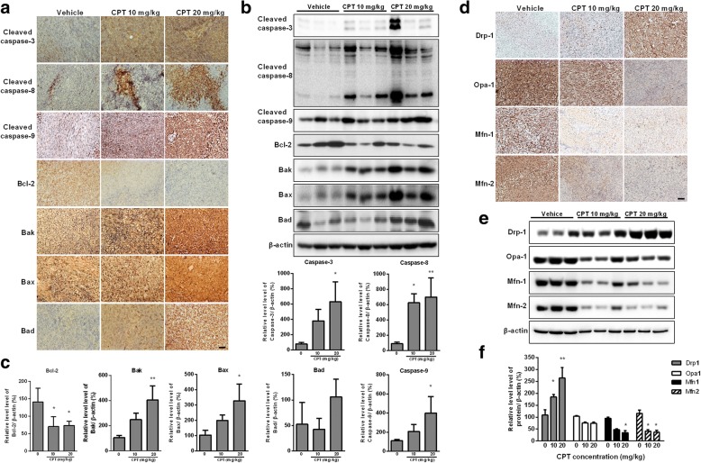
Fig. 7.
CPT activates caspase-mediated apoptosis and impaired balance of mitochondrial fission and fusion in vivo. 143B cell-derived tumors were developed in NOD/SCID mice and treated with CPT or vehicle for 45 days. Expressions of cleaved caspase 3, cleaved caspase 8, cleaved caspase 9, Bcl-2, Bak, Bax and Bad were examined by immunohistochemistry (a) and Western blotting (b). The quantification analysis in Figs. b is for Figs. (c). Expressions of Drp1, Opa1, Mfn1 and Mfn2 were examined by immunohistochemistry (d) and Western blotting (e). The quantification analysis in Figs. e is for Figs. (f). Ratios of each protein to β-actin were determined by densitometry. The results were expressed as the means ± SD from three independent experiments. *P < 0.05 and **P < 0.01, significantly different compared with control. Bar represents 500 μm.

