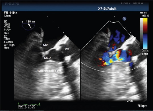
Figure 2.

A midesophageal aortic valve long-axis TEE view at 120° showing the hypertrophied interventricular septum predisposing a patient of severe AS to midcavity obstruction. TEE: Transesophageal echocardiography, AS: Aortic stenosis, MV: Mitral valve, Ao: Aorta
