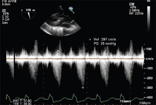
Figure 5.

A deep transgastric TEE view at 106° demonstrating a “dagger”-shaped continuous-wave Doppler profile across the LVOT with a characteristic late systolic peaking, in a case of post-AVR patient for predominant AS. TEE: Transesophageal echocardiography, LVOT: Left ventricular outflow tract, AVR: Aortic valve replacement, AS: Aortic stenosis
