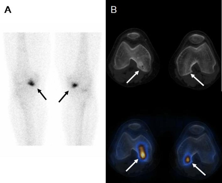
Figure 5.
Avascular necrosis. A 65-year-old male with a history of renal transplantation and long-term steroids treatment presented with bilateral knee pain. (A) Planar bone scintigraphy shows non-specific focal increased osteoblastic activity at the medial aspects of both knee joints (arrows). (B) SPECT/CT localises the osteoblastic foci to the sclerotic marrow lesions at the posterior aspects of both medial femoral condyles, which are more prominent on the right side (arrows).[ Reproduced with kind permission from Lu SJ et al; Value of SPECT/CT in evaluation of knee pain. Clin Nucl Med 2013; 38 (6): e258–6.035]

