Figure 2. .
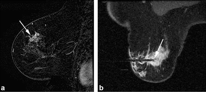
A 69-year-old female presented with palpable area of thickening in left superior breast with no correlate seen on mammography or sonography. (2A) Sagittal T1- post-contrast (subtraction) breast MRI image demonstrates clumped non-mass enhancement with segmental distribution at 12 o’clock position, corresponding to area of palpable finding (arrow). (2B) Axial T1 weighted post-contrast image obtained during MRI-guided vacuum-assisted needle biopsy confirms obturator position within suspicious lesion targeted for MRI-guided biopsy (arrow). Histopathology from VAB revealed ADH. Surgical excision was recommended and upgrade to DCIS, intermediate grade involving an area of approximately 2.5 cm, was revealed at segmental mastectomy. ADH, atypical ductal hyperplasia; DCIS,ductal carcinomain situ; VAB, vacuum-assisted biopsy.

