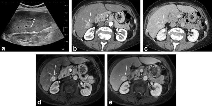
Figure 1.
HCC discovered during surveillance in a 56-year-old male with HCV-related cirrhosis. Systematic ultrasound depicted a mildly hypoechoic 25 mm subcapsular in Segment 6 (arrow in a). On contrast-enhanced CT (b, c) and gadoxetic-acid enhanced MRI (d, e) the lesion showed marked and homogeneous enhancement on arterial phase (b, d), and no washout on portal venous phase (c, e). The lesion was biopsied and proven to be a well-differentiated HCC. The patient underwent resection and remains recurrence-free at 23 months. HCC, hepatocellular carcinoma; HCV, hepatitis C virus.

