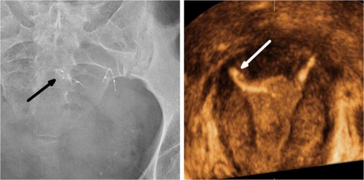
Figure 12.
A 45-year-old multiparous female who underwent a complicated Essure procedure because of poor visibility with 14 trailing coils visible after the deployment of the right insert. A history of angioedema after an intravenous urography prevented the use of hysterosalpingography. (a) A pelvic radiography showing a curled right insert (arrow). (b) 3D reconstructed coronal view of the uterus confirming the abnormal configuration of the insert (arrow) in the right cornua. 3D, three-dimensional.

