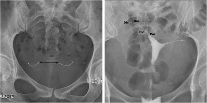
Figure 3.
Normal appearance of Essure birth control device on radiography (a) and hysterosalpingography (b). (a) Pelvic X-ray showing two symmetrical inserts, laying in the pelvic area without any abnormal configuration. Their radiomarkers are aligned and the distance between the proximal ends of the inserts is inferior to 4 cm. (b) Hysterosalpingogram showing correct placement of the two inserts with bilateral tubal occlusion. Both inserts have an intrauterine portion, an interstitial portion and a tubal portion. Contrast agent fills both cornual regions without progression into the fallopian tubes or intraperitoneal spillage. Pic/Dic, proximal and distal markers of the innercoil; Poc/Doc, proximal and distal markers of the outercoil.

