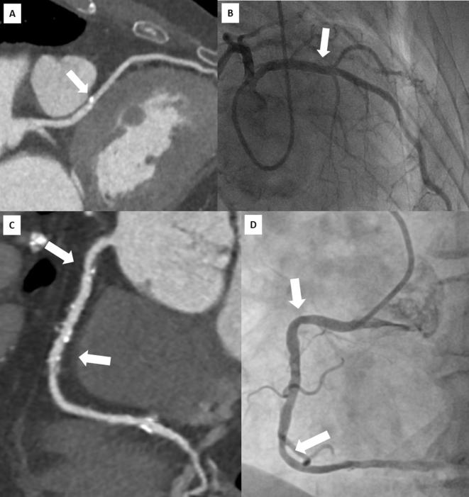
Figure 4.
Head-to-head comparison of CT multiplanar reconstruction (A) and ICA (B) showing a calcified plaque of proximal LAD (arrow) correctly ruled out as not significant at CT examination. Lower panels show a multiplanar reconstruction of a RCA with widespread atheromatous lesions (arrows) defined as “positive” by CT (C) but not confirmed during ICA (D).

