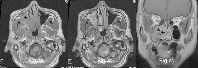
Figure 2.
Images in a 58-year-old male with OP of the right maxillary sinus (a) and (b), Right maxillary sinus has a well-demarcated and lobulated mass extending into right nasal tract, which reveals heterogeneously high signal (★) on axial T1- and isointense signal on T2 weighted MR images interspersing with multiple variable cystic changes of high T2 signal (+). (c) The focus exhibits heterogeneous enhancement with a typical “cerebriform” sign (*) on coronal fat-suppressed contrast-enhanced MR image. No enhancement of the cystic regions is noted (+).

