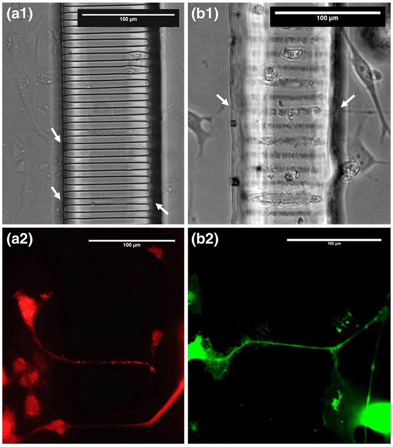
Fig. 7.

Cells within the Mμn System. MLO-Y4 osteocytes and B35 motor neurons adjacent to the nanochannel array construct. Phase contrast images of MLO-Y4 (a) and B35 (b) demonstrating cell phenotype and contact with channel array. Fluorescence images of Calcein AM stained cell MLO-Y4 (c) and B35 (d) demonstrating process extension across the array. Black arrows indicate process in-growth. Dashed lines indicate channel array boundaries
