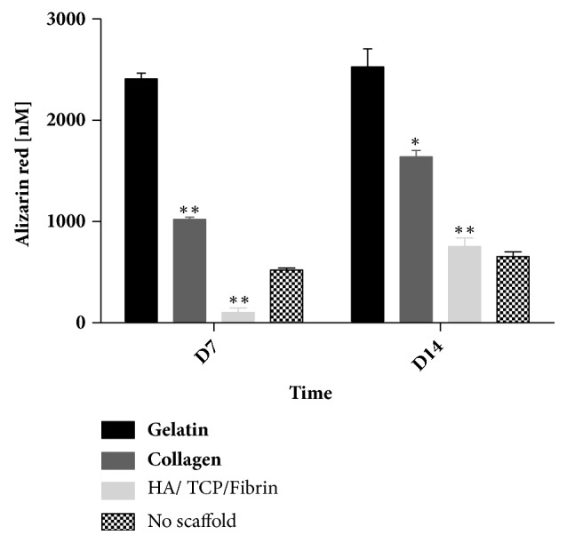
Figure 3.

Impact of scaffold type on calcium deposition by hMSSM-derived cells. Cells were seeded either alone or on collagen, gelatin, or HA/βTCP/Fibrin scaffold and cultivated in osteogenic differentiation medium for 14 days. Alizarin red quantification was used to assess calcium deposition. Each value represents a mean ± SEM for three independent experiments (n=3) each done in triplicate. ∗p<0.05; ∗∗p<0.01 vs. cells with gelatin scaffold (Student's t-test).
