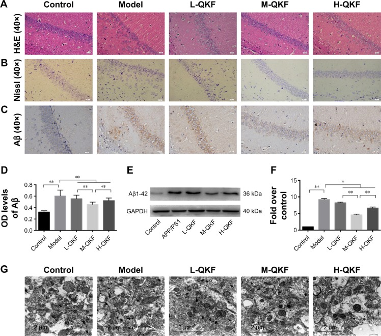
Figure 2.
QKF alleviate pathological degeneration in the brain of APP/PS1 mice.
Notes: (A) Nissl staining in CA1 region of the hippocampus (original magnification, ×400; scan bar, 20 µm). (B) Nissl staining in CA1 region of the hippocampus (original magnification, ×400; scan bar, 20 µm). (C) Immunohistochemistry for Aβ expression in the CA1 region of the hippocampus for each group (original magnification, ×400; scan bar, 20 µm). (D) Analysis of Aβ deposition levels by immunohistochemical staining. Values are expressed as the mean±SEM, n=30 per group. Significant differences between groups are indicated as *P<0.05 and **P<0.01. (E) WB for Aβ1-42 expression in the hippocampus of mice. (F) ODs indicative of Aβ1-42 protein expression. Values are expressed as mean±SEM, n=5 per group. Significant differences between groups are indicated as *P<0.05 and **P<0.01. (G) Ultrastructural observation of the CA1 area in the hippocampus of mice in each group using a transmission electron microscope (original magnification, ×18,500; scan bar, 2 µm). L-QKF, low-dose QKF group; M-QKF, middle-dose QKF group; H-QKF, high-dose QKF group.
Abbreviations: QKF, qingxin kaiqiao fang; Aβ, amyloid β; SEM, standard error of the mean; WB, Western blot.

