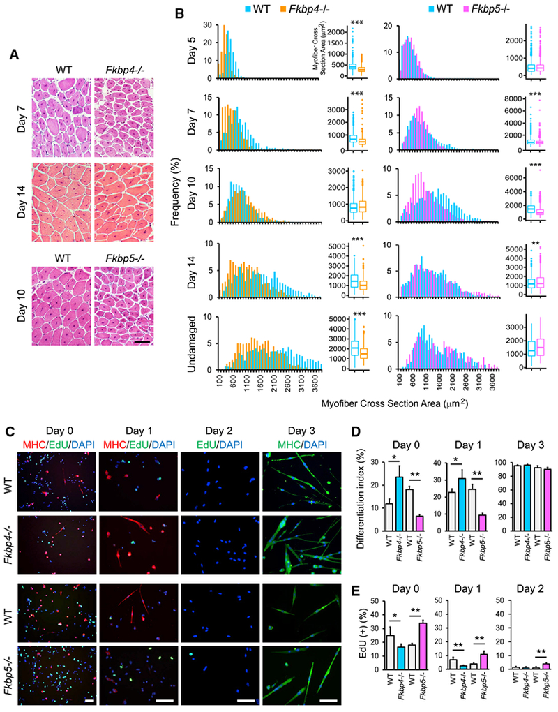
Figure 1. Regeneration of TA Muscles in Fkbp4−/− and Fkbp5−/− Mice and Differentiation of Their Myoblasts.

(A) H&E staining of TA muscle sections of KO and WT mice. Scale bar, 100 μm.
(B) Size distribution of regenerating myofibers, which contain centrally located nuclei, and undamaged myofibers stained with H&E. Randomly selected 500 myofibers from two mice each, totaling 1,000 myofibers for each mouse strain, were measured. *p < 0.01, **p < 0.001, and ***p < 0.0001 with Wilcoxon Rank Sum test with Bonferroni adjustment.
(C) Immunostaining of primary myoblasts prepared from WT and KO mice with antibodies against MHC. EdU uptake was also detected. DNA was counter stained with DAPI. Cells were induced to differentiate with 5% horse serum for one and three days. Scale bars, 50 μm.
(D) The differentiation index of primary myoblasts.
(E) EdU uptake of differentiating myoblasts.
*p < 0.05 and **p < 0.01 with Student’s t test in (D) and (E). Data are presented as mean + SD of technical triplicates in (D) and (E).
