Fig. 7.
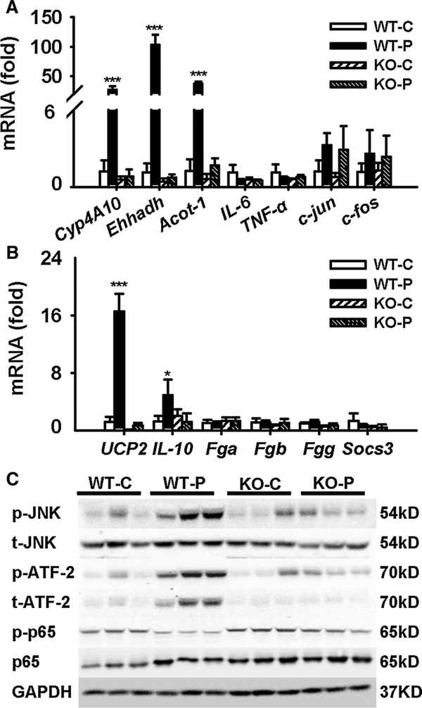
PFDA induced inflammation and anti-inflammation mediated by PPARα. a, b mRNA level of PPARα target genes, inflammation, apoptosis, necrosis-related factors, and STAT3 target genes. c Western blot of JNK and NFκB signaling in liver extracts. Data were from liver samples collected 5 days after PFDA treatment, and three of them were randomly selected for protein analysis. GAPDH was used as a loading control. The molecular weight was indicated at the left side of each band. Data were expressed as mean ± SD (n = 5; *p < 0.05; ***p < 0.001)

