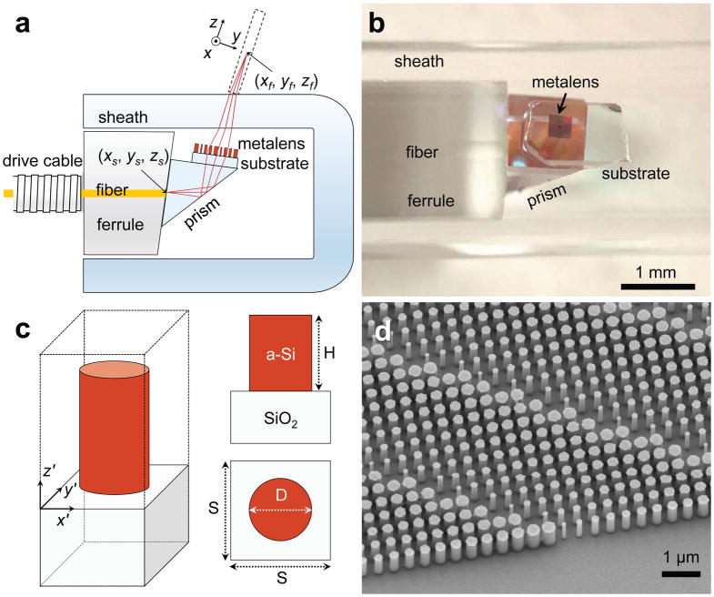
Figure 2. Nano-optic endoscope design and fabrication.
a, Schematic of the nano-optic endoscope. The metalens was designed to image a point source at (xs, ys, zs) to a diffraction-limited spot at (xf, yf, zf) with working distance WD = 0.5 mm. b, Photographic image of the distal end of the nano-optic endoscope. c, Schematic of an individual metalens building block consisting of an amorphous silicon (a-Si) nanopillar on a glass substrate. The nanopillars have height H = 750 nm and are arranged in a square lattice with unit cell size S = 400 nm. Phase imparted by a nanopillar is controlled by its diameter (D). d, Scanning electron micrograph image of a portion of a fabricated metalens.

