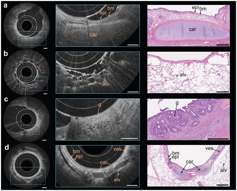
Figure 6. In vivo and ex vivo endoscopic OCT using the nano-optic endoscope.
a–d Endoscopic imaging of ex vivo human lung resections (a, b, c) and in vivo in the upper airways of sheep (d) using the nano-optic endoscope. Structural features of lung tissue were clearly visible in the magnified OCT images (centre column) including moderately scattering epithelium (epi), highly scattering basement membrane (bm), cartilage (car), blood vessel (ves), and alveoli (alv). Fine features, including the small irregular glands (g), the hallmark of adenocarcinoma, can be discerned. Representative histological images are provided in the right column. All scale bars are 500 μm.

