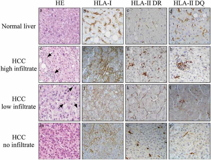
Figure 1.

HLA class I, but not HLA class II, is highly expressed on HCC tumor cells.
Immunohistochemical staining for both HLA class I and HLA class II in paraffin-embedded blocks of HCC tissue samples. The upper panels (a-d) show normal liver tissue with HLA class I and HLA class II expression (here assessed for both HLA-DR and HLA-DQ) confined to LSEC and KC cells. In contrast, the HCC tumor tissues, classified as having high infiltrate (panel e, arrowheads), low infiltrate (panel i, arrowheads), or no infiltrate (panel m), show strong membrane expression of HLA class I (panels f, j, n), but no expression HLA class II (panels g, h, k, l, o, p) in tumor cells. Original magnification X 400.
